Skin grafts are used to cover large wounds. When properly transplanted, they can integrate with the surrounding tissues and heal quite nicely.
While tissue engineering has enabled scientists to create “artificial skin” grafts, an autologous full-thickness graft remains the gold standard for this type of treatment. However, improper harvesting causes a significant drawback: inadequate re-epithelialization which leads to donor site morbidity.
This is a problem Christine Fuchs and her research team from the photomedicine and dermatology departments at the Massachusetts General Hospital and Harvard Medical School, Boston have been working on. In a study published in Science Advances, the team reports their innovative method of harvesting a full-thickness graft.
There’s a large magnet involved.
Micro skin grafts
According to the paper, current tissue engineering techniques cannot replicate the complex structure of natural skin.
“While there are ongoing efforts to increase the inclusion of various skin cell types into artificial skin constructs, there is currently no engineered skin product that can incorporate all the diverse cell populations/subpopulations or reproduce the intricate extracellular architecture of natural skin.”
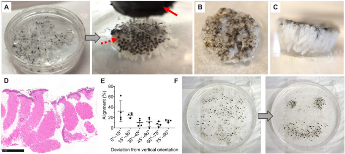
In an earlier study, the team came up with a technique that allows them to harvest natural full-thickness skin grafts. This “top-down” strategy involves punching out micro skin tissue columns (MSTCs). These are small enough so that the harvest site can heal easily. Furthermore, the MSTCs can be harvested from all over the body, introducing a much-needed diversity in the completed tissue.
However, the study showed that using such a tissue made from randomly harvested MSTCs delayed wound healing as the tissue went through a reorganization process between the epidermal and dermal cells.
Therefore, in the present study, they wanted to observe how the wound behaves when transplanted with a properly organized tissue.
They created did tissue by coating the donor site with a magnetic material made by blending a silicone-based wound adhesive with iron oxide. Using a magnet would keep the natural orientation of the dermal cells, controlling the tissue organization.
Fittingly, the team named this the MagneTEskin.
When testing in vivo on a porcine model, the researchers found that the tissue healed well during the first few days. It then degraded due to the incompatible extracellular binding materials used.
Although the attempt was unsatisfactory, this new technique is quite revolutionary. If perfected, the method could critically enhance wound care around the world.
Source: Science Advances
https://www.science.org/doi/10.1126/sciadv.abj0864


