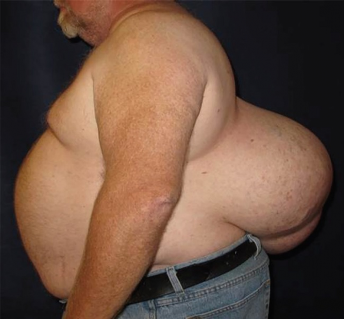
Male patient presented to the clinic with a 14 kg subcutaneous lipoma that had been present for 20 years.
A 54-year-old male patient presented to the clinic with a 14 kg subcutaneous lipoma that had been left untreated for 20 years. The patient’s past medical history was notable for hypertension. On examination a soft tissue growth was present on his lower back. According to the patient, the growth has been rapidly enlarging over the past three years. Moreover, had nearly doubled in size during this tenure.
This was the first time the patient visited a physician since childhood. At the time of presentation the patient did not complain of any pain or tenderness over the mass. Moreover, there was no history of fever, night sweats and weight loss. The rest of the physical examination did not reveal any significant findings excluding the large soft tissue mass over the patient’s lower back. The dimensions of the mass were measured to be 38 cm.
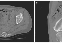
The patient was sent for evaluation to surgical oncology and radiation oncology services. The patient was advised a CT scan which showed a large (35 cm, x 38 cm x 17 cm), heterogeneous soft tissue mass. Based on findings of the radiological imaging, the differential diagnosis was narrowed down to teratoma and liposarcoma. Histopathological analysis of the biopsied mass showed fat necrosis with calcifications.
The rapidly growing mass was diagnosed as a benign giant lipoma
A 6 hour operation was performed to excise the 14 kg mass, 4 weeks after presentation. Similarly, after the patient was prepped and draped, the skin overlying the central portion of the tumour was shaved and harvested as split thickness skin grafts.

In addition, an incision was made in the skin overlying the tumour in the area outside the skin graft donor sites and significant flaps were preserved in all dimensions to permit primary closure.

The tumour was dissected off and several, large variceal vessels feeding the tumour were ligated. The dissected specimen was sent for frozen section analysis. The findings were consistent with the diagnosis of a lipoma, confirming the initial diagnosis.
Moreover, the preserved skin flaps were used to primarily close the defect which measured more than 200 cm x 40 cm.

The skin flaps were de-epithelialized and were overlapped to achieve a multilayer closure of the back wound with the intention to obliterate most of the deadspace. Additionally, prior to the final closure, two subcutaneous flaps were placed.
The patient recovered well without any postoperative complications. The patient was discharged after an uneventful stay at the hospital in good condition. The drains of the patient were sequentially removed and the incision line healed without any discomfort.
The patient showed no evidence of infection or recurrent at his 6-month follow-up.
References
Untreated for 20 Years: A 14 Kilogram Subcutaneous Lipoma https://www.ncbi.nlm.nih.gov/pmc/articles/PMC6290305/


