- Desmoid tumours are locally aggressive tumours with no potential for metastasis.
- The tumours present as slow-growing, non-specific solid masses, making it difficult to distinguish from other soft tissue tumours.
- Sporadic desmoid tumours can be found at any site of the body.
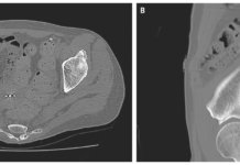
A 52-year-old woman presented was incidentally detected with an intraabdominal desmoid tumour while she was undergoing a postoperative follow-up CT for chondrosarcoma of the rib. She family medical history was unremarkable. Laboratory tests showed serum biochemistry results within normal limits. An abdominal CT was further performed which showed a defined a well-defined cystic mass, measuring 20 x 18 mm with a solid component. The tumour was visible adjacent to the stomach and the pancreas. However, there was no sign of invasive growth.
For further evaluation, an MRI was performed which showed a cystic mass on T2-weighted images with high intesnity and T1-weighted images on low intensity. Endoscopic ultrasound showed an 18 x 3 mm round cystic mass and a heterogenous nodular lesion on the side of the serosa of the stomach. No local lesions were evident on the mucosal surface of the stomach on esophago-gastric endoscopy. The findings were consistent with an intraabdominal tumour. However, it was difficult to determine a specific diagnosis.
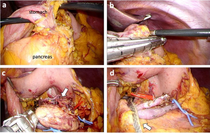
Treatment included laparoscopic resection of the tumour. The tumour was confirmed at the superior border of the pancreas body, adhering to the posterior wall of the stomach. A distal pancreatectomy was done to preserve the spleen. A complete laparoscopic excision was done, preserving the spleen and splenic vessels. Intraoperative blood loss was 15 mL. The procedure time was 279 minutes.
The patient’s postoperative recovery was uneventful and was discharged healthy.
References
Laparoscopic spleen-preserving distal pancreatectomy for a solid-cystic intraabdominal desmoid tumor at a gastro-pancreatic lesion: a case report https://bmcsurg.biomedcentral.com/articles/10.1186/s12893-020-0691-5


