Case Report
Femoral-facial syndrome (FFS, OMIM 134780) is an uncommon syndrome with various congenital defects. Affecting the femur, cranium, mid-face, and extremities. Its prevalence has been estimated to be less than 1/1,000,000. Although an etiological connection with maternal diabetes has been identified, it is rare.
The genetic transmission of this illness is linked to the placements of genes on autosomes (dominant) in some families, although no specific gene mutation has been discovered.
The primary characteristics of this condition are bilateral femoral aplasia/hypoplasia and cranial-facial dysmorphism; however, a wide spectrum of congenital anomalies was documented in 1975. Micrognathia, low-set ears, broad nose, long philtrum, flat philtrum, cleft palate, thin upper lip, upward-slanting eyelids, and craniosynostosis are some of the craniofacial characteristics. Other systemic irregularities may potentially be linked to FFS.
Congenital cardiac disorders, like septal abnormalities and patent ductus arteriosus, are the most frequent in FFS. FFS may be related to chest abnormalities, agenesis, or a polycystic kidney. Other skeletal manifestations of FFS include scoliosis, long bone synostosis, talipes equinovarus, macrodactyly, and polydactyly.
Case Presentation
A one-day-old Black Bantu African preterm female baby weighing 1000 g was born spontaneously vaginally to a Gravida 3, Living 2 mother. She was admitted to the neonatal critical care unit shortly after birth due to several congenital abnormalities and moderate respiratory distress.
The mother is 32 years old, married, but not related to her husband. Due to placental abruption, the mother did not seek medical assistance until the early third trimester of her pregnancy. The first prenatal ultrasound showed significant oligohydramnios. She did not have diabetes (random blood glucose was 5.1 mmol/L) and did not take folic acid treatment during pregnancy.
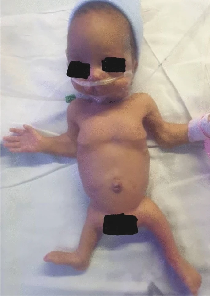
The baby was alert upon admission, with modest symptoms of respiratory distress syndrome. She exhibited dysmorphic traits like microcephaly (head circumference < 10%), large occiput, low-set ears, right cleft palate and lip, micrognathia, bilateral elbow contracture, and shortened lower limbs.
The baby weighed 1000 g at birth, measured 40 cm long, and had an occipitofrontal circumference of 30 cm.
Investigations
Skeletal X-rays revealed rib flattening, right femur aplasia, and left femur hypoplasia. The laboratory workout included complete blood counts, c-reactive protein, creatinine, blood urea, and electrolytes, all of which were normal. The cranial ultrasound was normal, but echocardiography indicated a tiny patent ductus arteriosus.
Treatment
The infant was kept on respiratory assistance and given empirical antibiotics. An orthopedic surgeon, an ENT surgeon, and a physical therapist were involved. The cleft palate will be surgically repaired at the age of six months. Functional therapy is planned before the patient begins walking. Limb lengthening surgery is not currently performed in our context.
Discussion
The clinical diagnosis of FFS has shifted from femoral hypoplasia plus one or more prominent craniofacial characteristics to femoral hypoplasia plus two or more facial deformities.
The etiology of FFS is unknown, and the majority of cases occur randomly. Maternal hyperglycemia is thought to be a teratogenic exposure, accounting for 38% of occurrences of FFS. Other potential exposures have been identified, including prenatal medications, maternal illnesses, and oligohydramnios. In this case, the mother did not develop hyperglycemia during the third trimester till delivery. There was no family history of congenital defects, but she scheduled antenatal care after 28 weeks of gestation.
Caudal dysgenesis in an infant born to a diabetic mother is caused by mesodermal insufficiency. The etiology of caudal regression syndrome and FFS is somewhat similar. During embryogenesis, the lateral plate mesoderm proliferates and develops into chondrocytes, which thereafter form the femur. The mother was not tested earlier in the pregnancy, but she was euglycemic when she scheduled prenatal treatment at 28 weeks.


