Rare case of papillary thyroid cancer
This article describes the case of a 55-year-old women diagnosed with a rare case of multifocal papillary thyroid cancer in bilateral thyroid cysts. The patient presented to the emergency with a neck swelling with a history of a few days. However, she did not complaint of any compressive symptoms. On clinical examination, a 4 cm left smooth thyroid nodule was notable.
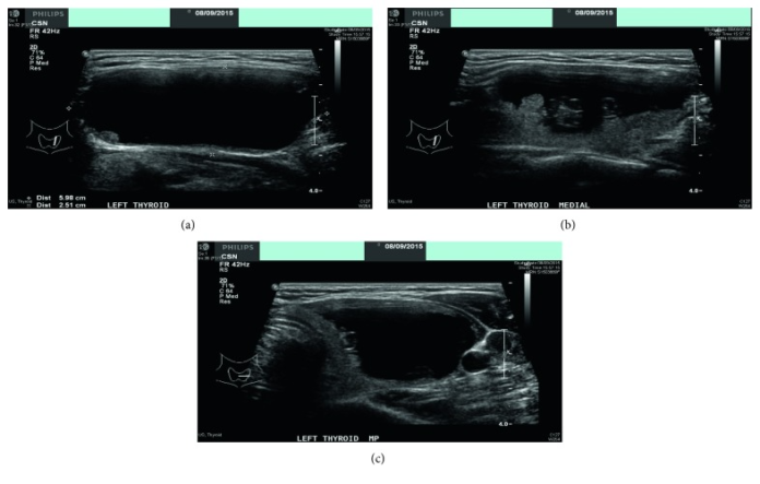
Doctors advised a thyroid function test which showed normal results. The ultrasound scan showed a cystic nodule measuring 5.9 x 3.8 cm with a small solid content. Furthermore, the nodule was smooth on palpation and did not show any increased vascularity. The right lobe showed 2 simple cysts which looked benign. The cysts measured 0.4 by 0.4 cm, 0.3 by 0.3 cm. However, there were no signs of cervical lymphadenopathy.
Doctors further advised an ultrasound-guided FNA. The aspirate was suggestive of cyst contents because of the aspirate reported. Considering the size of the left cystic nodule, the initial treatment plan included a left hemithyroidectomy. Doctors counselled the patient for this treatment approach. The patient’s recovery period after the procedure was uneventful. The final histology did not show any extrathyroidal extension or lymphatic invasion. Although, the patient opted for a complete right thyroidectomy to minimise any risk of disease occurrence. In addition, to optimise postoperative surveillance.
The lesion was biopsied which showed 2 foci of 3 mm each, confirming the diagnosis of papillary thyroid cancer. A postoperative radioiodine scan was performed which did not show any distal metastasis. She was started on thyroxine replacement. 1 year follow-up ultrasound scan and thyroglobin marker did not reveal any signs of cancer occurrence.
References
A Rare Case of Multifocal Papillary Thyroid Cancer in Bilateral Thyroid Cysts https://www.ncbi.nlm.nih.gov/pmc/articles/PMC5932504/


