35-year-old female patient with history of chronic asthma presented with cough and shortness of breath, diagnosed with tracheal adenoid carcinoma.
A 35-year-old female patient presented with a 1-year history of cough and shortness of breath. The clinical and investigation findings were consistent with tracheal adenoid cystic carcinoma.
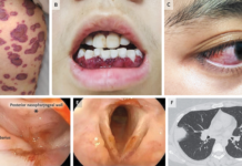
Doctors were currently treating her for asthma. However, she did not respond to treatment and developed hemoptysis. CT findings were remarkable of a soft tissue mass arising from the posterior and lateral wall of the trachea with mediastinum extension. Bronchoscopy revealed a tracheal mass 3 cm above carina with invasion of the lateral wall. The mass was biopsied with bronchial washing.
Histopathological analysis
The bronchial washing specimen was used for preparing alcohol-fixed smearing. Including, staining of smears with modified Papanicolaou method. Similarly, cytology showed hypercellular smears composed of loosely cohesive sheets. Dispersed cells and three dimensional clusters. The size of the cells was relatively small with round nuclei, small nucleoli and a scant cytoplasm. The smears showed different sizes of accellular hyaline materials with globule formation. In addition, the tumour cells were also encapsulating them.

In addition, a cell block was prepared with thrombin method. The cell block showed nests and strands with tubular-like structures and a cribriform pattern containing homogenous acidophilic materials.

Biopsy findings were consistent with the diagnosis of adenoid cystic carcinoma. Similarly, the biopsy showed bronchial mucosa with infiltrative neoplastic lesions composed of tubular and cribriform structures with acidophilic materials. The initial treatment plan was referring her for resection surgery. However, because resecting the tumour completely was not possible, she was advised radiotherapy.
References
Tracheal Adenoid Cystic Carcinoma Presented with Chronic Asthma Diagnosed by Bronchial Washing Cytology https://www.ncbi.nlm.nih.gov/pmc/articles/PMC6988659/


