2-year-old girl presented with symptoms of fever, irritability and a 7-day history of maculopapular rash.
A 2-year-old girl was diagnosed with coronary artery aneurysm and thrombosis secondary to Kawasaki disease. The girl presented with symptoms of fever, irritability and a maculopapular rash that had a history of 7 days.
Physical examination showed that the patient was febrile and tachycardic. Cervical lymphadenopathy, palmer and planter erythema was also present in association with bilateral conjunctival injection and congestion of oral mucosa and throat. However, the patient’s systemic examination was normal. In addition, hemoglobin was 11.2 g/dl, total white blood cell count was 16,100/mm3 with ESR of 60 mm at the end of 1 hour and platelets were 4.2 × 10⁵/mm3.
The clinical findings and investigations were consistent with the diagnosis of Kawasaki disease.
Coronary artery aneurysm
A coronary artery aneurysm is defined as a coronary artery dilation that exceeds the diameter of the largest coronary artery by 1.5 times. A giant coronary aneurysm is an aneurysm with a diameter larger than 20 mm.
Kawasaki disease
Kawasaki disease is a common cause of heart disease in children. It causes inflammation in arteries, veins and capillaries. Moreover, it affects the lymph nodes and causes symptoms in the nose, mouth and throat.
Treatment plan
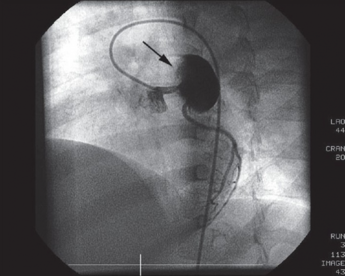
The patient was started on intravenous immunoglobulins (IVIg) and a high-dose of aspirin. After putting her on the medications, the symptoms improved significantly. An echocardiography was done when the patient was first admitted. Results showed a giant aneurysm measuring around 18 mm, of the proximal left anterior descending artery with thrombus.
She was further prescribed warfarin with the dose adjusted to maintain INR of about 2.5. 2 months later, a follow-up echocardiogram was performed which showed an increase in the size of the aneurysm, measuring 22 mm. However, the thrombus had completely resolved. Although, the patient remained asymptomatic throughout her follow-up period.
The patient was advised a coronary angiography given the progressive increase in the size of the aneurysm, to delineate the coronary anatomy. The coronary angiography showed a giant aneurysm of 25 mm just after the origin of LAD with swirling of contrast within the aneurysm. In addition to a faint and delayed opacification the the distal LAD. The left main coronary artery (LMCA) and left circumflex coronary artery (LCX) were normal. The right coronary artery was also normal, supplying collaterals to the LAD.
References
Giant coronary artery aneurysm with a thrombus secondary to Kawasaki disease https://www.ncbi.nlm.nih.gov/pmc/articles/PMC2840734/


