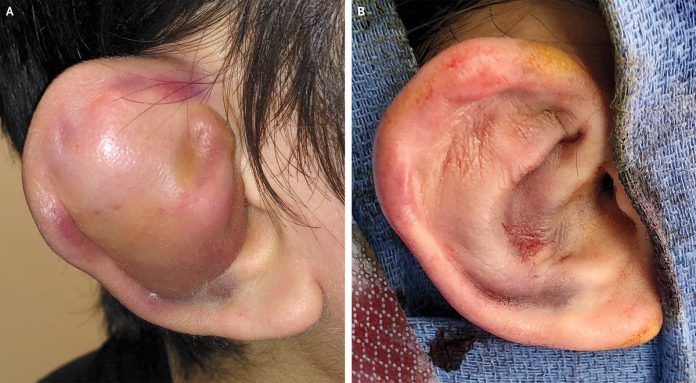
Auricular hematoma:
- Accumulated blood underneath the perichondrium of the ear.
- Typically caused by trauma.
- It is imperative to examine the external ear and the tympanic membrane with an otoscope in patients with auricular hematoma.
- Drainage is an appropriate management strategy, especially within the first 48 hours.
- If left untreated or not treated adequately, or if repeated trauma occurs, it may lead to an auricular deformity called the “cauliflower ear.”
- Post-drainage, it is best for the patient to limit physical activity for the next 10 to 14 days and avoid contact sports for 1 to 2 weeks.
A 17-year-old boy was brought to the emergency department with complaints of progressive swelling and bruising of the right ear. The patient was a known case of autism spectrum disorder. He had a history of multiple episodes of hitting his head on the floor, consequently injuring himself; a similar episode had occurred one day before the presentation, according to the parents.
Physical Examination:
General examination revealed an alert, healthy teenager who was comfortably sitting and was not in any distress. Just above his left eyebrow, there was a visible minor laceration. Inspection of the right ear showed marked swelling with overlying ecchymosis and a loss of cartilaginous landmarks. In palpation, the swelling was fluctuant.
The findings of the physical examination of the right ear were consistent with the diagnosis of an auricular hematoma (Panel A).
Management:
It was decided to perform an urgent incision and drainage of the auricular hematoma; therefore, the patient was taken to the operating room.
Urgent drainage was performed, followed by (Panel B) shaping the ear’s contours with several bolsters. The surgery was uneventful. Post-drainage, bolster dressing is imperative to prevent the reaccumulation of hematoma/fluid collection in the potential space.
Postoperatively, the bolsters were removed on the third day. A month after the procedure, the patient had two more self-injurious episodes, subsequently leading to the recurrence of right auricular hematoma.
Twice again, the patient underwent drainage and bolster-application.
At the 6-month follow-up, the patient had developed thickening of the right auricular cartilage along with contour loss.
References:
Ashley L. Miller, M. a. (2020, November 05). Auricular Hematoma. Retrieved from The New England Journal of Medicine: https://www.nejm.org/doi/full/10.1056/NEJMicm2004765
Krogmann RJ, King KC. Auricular Hematoma. [Updated 2020 Aug 8]. In: StatPearls [Internet]. Treasure Island (FL): StatPearls Publishing; 2020 Jan-. Available from: https://www.ncbi.nlm.nih.gov/books/NBK531499/


