
A case of pleural plaques because of secondhand exposure to asbestos
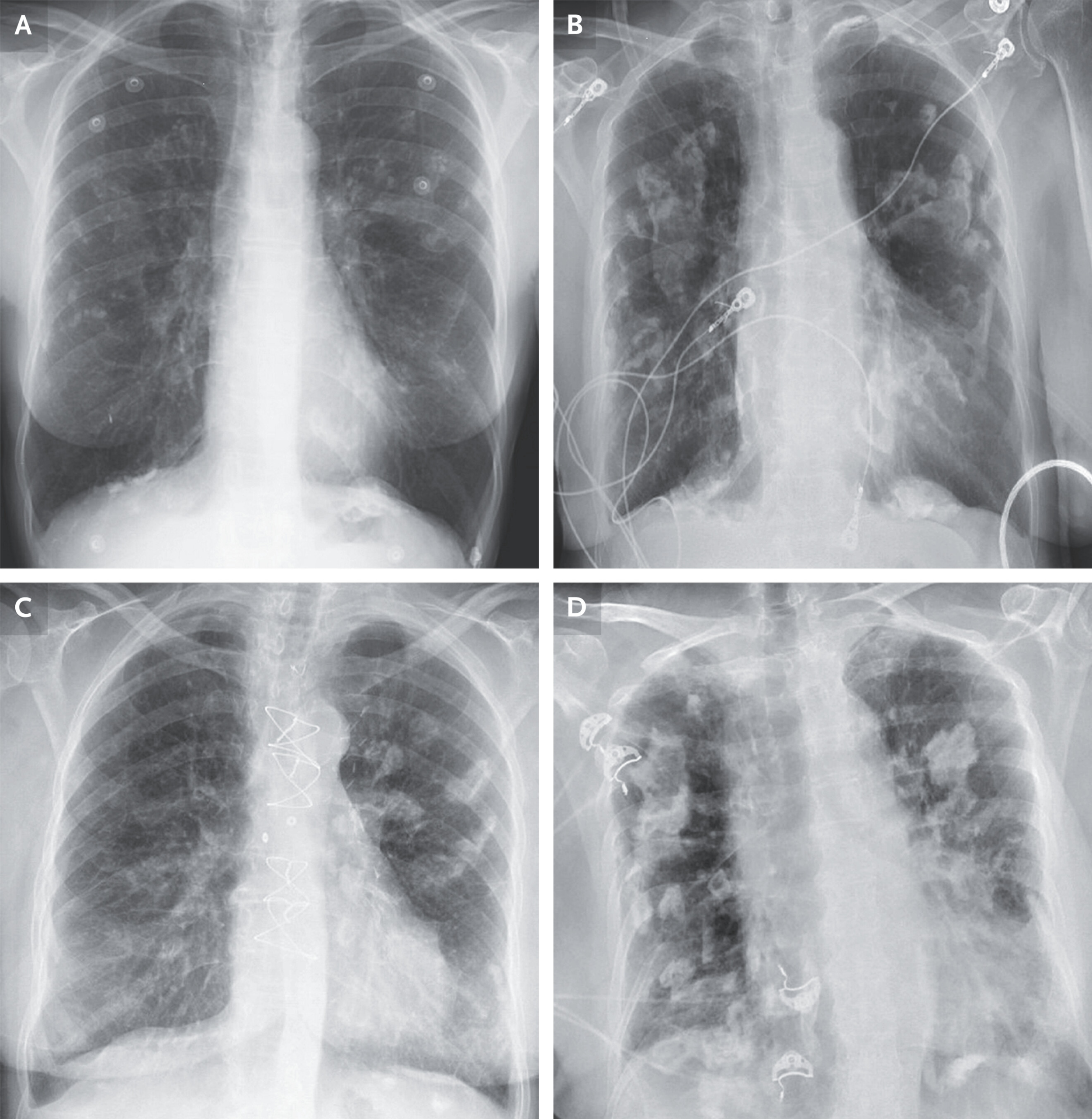
An 80-year-old woman was found to have pleural plaques on a routine chest radiograph. Although she had no respiratory symptoms at the time of diagnosis, the imaging findings prompted further clinical inquiry. When her medical history was explored in more detail, a significant childhood exposure emerged: she recalled playing with her father when he returned home from work, his clothing often covered in fine, “snow-like” dust particles. This seemingly innocent memory provided an important clue to a long-standing occupational exposure that likely shaped her current diagnosis.
Pleural plaques are areas of localized thickening of the pleura, the thin membrane surrounding the lungs and lining the chest wall. They are considered the most common manifestation of prior asbestos exposure and serve as a marker of environmental or occupational contact with asbestos fibers. Importantly, pleural plaques themselves are usually benign and do not cause symptoms such as shortness of breath or chest pain. However, their presence is clinically significant because they indicate prior exposure to a known carcinogen.
Asbestos is a naturally occurring fibrous mineral that was widely used throughout the 20th century in construction, insulation, shipbuilding, and automotive industries due to its heat resistance and durability. Workers in these industries were frequently exposed to airborne asbestos fibers, which could cling to clothing, skin, and hair. Family members of exposed workers were also at risk through secondary or “para-occupational” exposure—exactly the type of exposure described in this case, where the patient’s father unknowingly brought asbestos dust home on his work clothes.
Once inhaled, asbestos fibers can become lodged in the pleura and lungs. Over time, they induce chronic inflammation and fibrotic changes. The pleura is particularly susceptible to plaque formation, especially along the parietal pleura on the chest wall and diaphragm. These plaques typically contain dense collagen and may calcify, making them visible on imaging studies such as chest radiography or computed tomography (CT) scans.
Radiographically, pleural plaques appear as discrete, well-defined areas of thickening, often with a characteristic “holly leaf” or calcified appearance. They are usually bilateral but may be asymmetrical. While chest X-rays can detect larger or calcified plaques, CT imaging is more sensitive and can identify smaller or non-calcified lesions that may be missed on plain radiographs.
Although pleural plaques themselves are not malignant and do not typically impair lung function, they are important because they signal prior asbestos exposure. This exposure is associated with several serious conditions, including asbestosis (a diffuse interstitial lung fibrosis), lung cancer, and malignant mesothelioma, a rare and aggressive cancer of the pleura. According to major health authorities such as the World Health Organization (WHO) and the U.S. Environmental Protection Agency (EPA), there is no safe level of asbestos exposure, and even limited or indirect exposure can carry long-term health risks.
The latency period between asbestos exposure and development of pleural disease can be extremely long, often ranging from 20 to 50 years. This explains why individuals like this patient, who had exposure in childhood, may only present with radiologic findings many decades later in adulthood or old age. In many cases, patients are unaware of their exposure history until careful questioning reveals indirect contact, such as household exposure from a family member working in a high-risk occupation.
In this case, the description of “snow-like particles” on the father’s clothing is highly characteristic of asbestos dust, which was commonly described in this way by family members of exposed workers. During the mid-20th century, before strict regulations were implemented, asbestos exposure in industrial settings was widespread. Even today, older buildings undergoing renovation or demolition can still release asbestos fibers into the environment if not properly managed.
The diagnosis of pleural plaques is usually made incidentally during imaging performed for other reasons. Once identified, further evaluation focuses on assessing the extent of asbestos exposure and screening for associated complications. Pulmonary function tests are often normal in patients with isolated pleural plaques, but they may be performed to establish a baseline and detect any early restrictive changes.
Management of pleural plaques themselves is generally conservative, as they do not require treatment. The primary clinical focus is on surveillance and prevention. Patients are advised to avoid further asbestos exposure and to stop smoking if applicable, as smoking significantly increases the risk of asbestos-related lung cancer. In some cases, periodic follow-up with imaging or pulmonary assessment may be recommended, particularly if there are additional risk factors or symptoms.
This case highlights the importance of taking a detailed environmental and occupational history, even when the exposure occurred indirectly or many decades earlier. It also underscores how seemingly harmless childhood memories can provide critical diagnostic clues in adult medicine. The recognition of pleural plaques serves not only as a radiologic finding but also as a marker of past exposure to a substance with well-established long-term health risks.
In summary, pleural plaques in this elderly patient reflect prior asbestos exposure, most likely acquired indirectly during childhood through contact with her father’s contaminated work clothing. While the plaques themselves are benign, their presence carries important implications for long-term health surveillance. This case serves as a reminder of the enduring legacy of asbestos exposure and the importance of occupational and environmental history in clinical diagnosis.
Source: NEJM