Case Report
Ambiguous genitalia is a condition caused by a sexual development abnormality, resulting in congenital defects in which the external genitalia do not mirror typical male or female traits.
Disorders of sex development DSDs are defined as aberrant development of chromosomal, gonadal, or anatomical sex. The overall incidence of ambiguous genitalia is about one in every 4500 births, with moderate variants of male under-virilization or female virilization occurring in about 2% of live births. Most ambiguous genitalia can be diagnosed at birth due to their vague outward traits.
Ambiguous genitalia in genetic females are frequently produced by disorders such as congenital adrenal hyperplasia (CAH), exposure to drugs with androgenic effects during pregnancy, or the presence of a male hormone-producing tumor in the fetus or mother.
In genetic males, potential causes include Leydig cell aplasia, 5-alpha-reductase deficiency, androgen insensitivity syndrome, or exposure to estrogenic drugs during pregnancy. Ambiguous genitalia can appear in DSD 46 and XY situations to variable degrees. Approximately half of underused boys are not given a formal diagnosis. According to studies, these newborns have lower birth weights than those with an established underlying cause. In addition, hypospadias has been associated with intrauterine growth restriction.
Case Presentation
On December 4, in a city in Iran, a 35-year-old rural Iranian lady, gravida 1, at 37 weeks of pregnancy (based on the first day of her last menstrual period), was admitted due to intrauterine growth limitation. A fter early preparations and mother preparation, a cesarean section was conducted.
The newborn weighed 1900 g, was 32 cm tall, had an Apgar score of 9 and 10. And showed evidence of ambiguous genitalia. The newborn’s parents had no kinship. And had no history of specific diseases, drug use, or allergies, nor did they have a history of a newborn with ambiguous genitalia or other sexual development abnormalities.
The mother had undergone conventional prenatal treatment. It is worth noting that during the first-trimester ultrasound, the fetus’ gender was reported as ambiguous genitalia. As a result, at 15 weeks of pregnancy, the mother had an amniocentesis to do a karyotype. That revealed the fetus’s genotype as 46XY, inv (9) (p12q13), with a normal chromosomal count. And a pericentric inversion of Chromosome 9. The phenotypic anomaly was caused by the inversion of Chromosome 9.
Examination
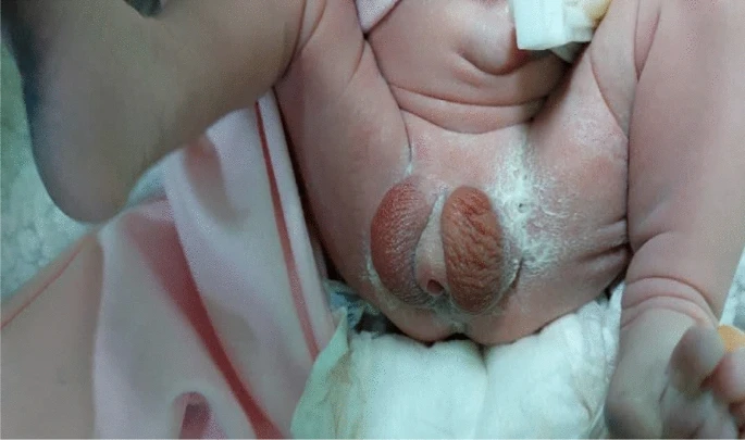
During a pediatrician’s physical examination of the newborn, a wrinkled scrotum resembling labia, severe hypospadias, and a micropenis were discovered. Due to the IUGR, the newborn was admitted to the neonatal intensive care unit (NICU). The patient’s electrolytes and glucose levels were within normal range. The newborn’s testosterone (T) and dihydrotestosterone (DHT) levels, assessed two days after delivery, were within normal range. The scan revealed no evidence of female genitalia. Further diagnostic tests at 2 months suggested a probable diagnosis.
Discussion
During the development and formation of fetal sex and external genitalia, chromosomal structure, gonads, and enzymes all play important roles. During the embryonic period, at the moment of zygote development, a primordial gonad develops, with cells. That have equal ability to differentiate into testes or ovaries. The gonads are where the first step of sexual development takes place, and chromosomal structure influences this stage.
In chromosomal configuration XY, the primordial gonad develops into testes, while in XX, it develops into ovaries. Gonadal differentiation into testes is complete by week 8 of gestation. If this pattern is not followed, the primordial gonad will naturally grow into an ovary by week 12 of gestation, unaffected by the HY-antigen (HY-AG).
Sexual ambiguity, also known as ambiguous genitalia in humans, is a condition in which the morphology of the gonads and the external genitalia do not match or coordinate. Sexual ambiguity is divided into two categories: real hermaphroditism and pseudohermaphroditism. True hermaphroditism develops when a person possesses both testicular and ovarian tissues. These patients’ external genitalia show varying degrees of intermediate masculinization and feminization. During adolescence, masculine and feminine secondary sexual characteristics emerge in various forms. Approximately two-thirds of people with this illness have a 46,XX karyotype, one-tenth have a 46,XY karyotype, and the rest have chromosomal mosaicism.
Pseudohermaphroditism is classified into two types: female and male pseudohermaphroditism. Female pseudohermaphroditism shares some male characteristics and is categorized into several groups, each with its own enzyme deficit and clinical symptoms. The most prevalent type is congenital adrenal hyperplasia, which is caused by 21-hydroxylase deficiency and is the major cause of sexual ambiguity in newborns. Male pseudohermaphroditism occurs when the individual has only testicular tissue, yet the external genitalia are feminized or incompletely masculinized.
Conclusion
Genital ambiguity is the most severe disorder on the DSD spectrum, and it is treated as a medical emergency. It is an uncommon condition with a contested global prevalence. Depending on the source, DSD might have long-term consequences, affecting not just sexual differentiation but also pubertal development and fertility in adults.
This report presents a healthy live delivery from a couple with no history of sickness, medication usage, or consanguinity, in which the newborn boy has ambiguous genitalia and a karyotype with inv (9, p12q13). Pericentric inversion of Chromosome 9 is a common chromosomal abnormality to consider in the differential diagnosis of ambiguous genitalia.


