A 21 year old female patient presented to the hospital with a complaint of swelling on the right neck. According to her, the swelling was 2 years old. However, it had not caused any significant symptoms. The patient also denied any family history of similar swellings or lesions.
Examination of the Swelling on the right neck and Investigations
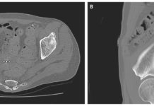
On examination, the swelling was found to be a non-pulsatile mass. Doctors concluded the mass has a smooth surface and mobile nature. It moved vertically but not horizontally. The mass was not associated with any compression symptoms. Moreover, pressing the mass did not elicit any cardiovascular change. Doctors did not find much information on history owing to the asymptomatic presentation and an insignificant past medical and family history. Thus, they proceeded with radiological investigations to reach a diagnosis. Ultrasound revealed a hypoechoic lesion very close to the carotid artery.
No evidence was found of cystic lesions or calcification. Interestingly, they observed that the mass was attached to the vagus nerve on both its proximal and distal parts.
Swelling on the Right Neck: Diagnosis?
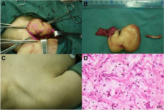
Doctors decided to remove the mass through surgery. Intraoperatively, they found the mass closely attached to the right vagus nerve just as indicated on the ultrasound preoperatively. However, pressing the mass did not elicit any cardiorespiratory changes. Thus, doctors resected the mass completely and then sent it for pathological examination. On histopathology, the mass was found to be a vagal neurofibroma. It was positive for GFAP and S100 proteins.
Doctors also noticed a small swelling on the left side of the neck. However, it was very small and asymptomatic. So, doctors decided not to operate on it. Overall, there were no intraoperative or postoperative complications while resecting the mass causing swelling on the right neck. Doctors also called the patient for a follow-up 8 months later where they did not observe any signs of recurrence.
Neurofibromatosis
Neurofibromatosis type 1 (NF1) is an inherited disorder that develops due to mutations in the NFT gene. It presents as skin lesions and swellings (neurofibromas) in association with a nerve (in our case, swelling on the right neck associated with the right vagus).
Neurofibromas associated with the vagus nerve and then involving them bilaterally are very rare. When present, they usually result in manifestations related to compression of the vagus nerve and surrounding structures. Thus, the common complaints are dysphagia, hoarseness of voice and cardiac changes like bradycardia. However, our patient did not have any of these.
Diagnosis
Preoperative diagnosis of neurofibromas, although critical for a surgical plan, remains a challenge. The list of differentials, in this case, includes an enlarged lymph node, a lipoma, a carotid body tumour, schwannoma and other tumours arising from nerves. The clinical presentation helps a lot in making the final diagnosis. But since our case was completely asymptomatic, the diagnosis was fairly difficult.
Some characteristics unique to particular tumours also help to narrow down the differentials. For example, schwannomas are usually isolated and unilateral, enlarged lymph nodes present with signs of tenderness and fever and carotid body tumours are associated with high blood pressure and deteriorating vision. Moreover, the closest differential schwannoma comes with cystic changes and remains completely resectable. Whereas, neurofibromas intertwine with the involved nerve and are very difficult to operate upon.
Management
The definitive treatment for such masses, especially when they are symptomatic, remains surgery. Complete resection along with strict follow-up for any complications and recurrence remains a standing operating protocol in this case.
As for our patient, doctors completely resected the mass on the right side. However, they found another mass on the left side although very small and asymptomatic. They decided not to operate upon it and monitor it for any changes instead. The patient was called up for a follow-up session after 8 months where he displayed good health with no signs of recurrence or any complications.


