Case of Mohs surgery for an extensive forehead squamous cell carcinoma!

0

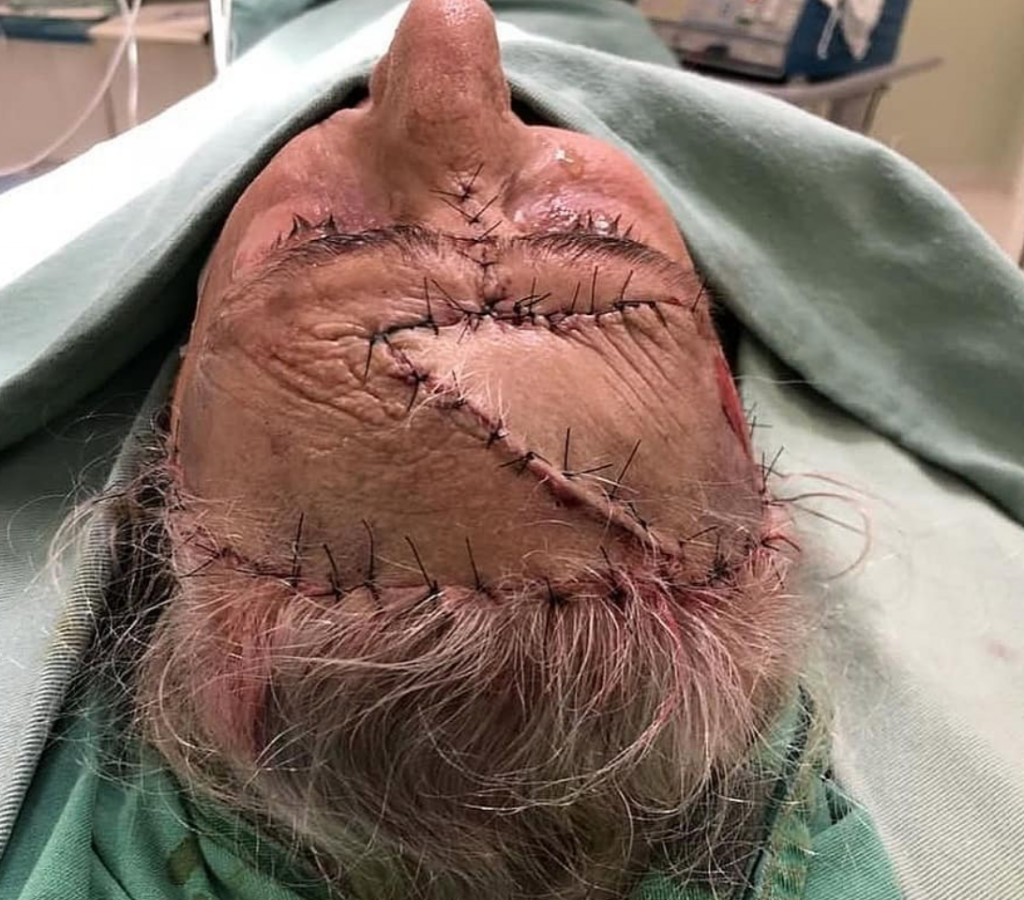
This patient underwent a Mohs micrographic surgery to treat her condition, which involves a sequential removal of thin skin layers with microscopic inspection to confirm that the margins have been cleared of malignant tissue.

This technique has the highest cure rate for basal cell carcinomas and in smaller lesions it provides the least disruption to surrounding tissues, making it ideal for delicate areas such as the perioral region, nose, lips, ears and in this case, scalp. Flap surgery, shown in last two photos followed shortly after to cover the defect.

Squamous cell carcinoma is a type of skin cancer that arise as a result of uncontrolled growth of the squamous cells in the epidermis layer of the skin and is suspected in patients with a rough, scaly nodule or non-healing, painless ulcer that develops in the setting of a scar or chronic inflammatory lesion. Risk factors are sun and radiation exposure, chronic wounds, burns, or scars. It is rapid growing and invasive, thus diagnosis is made by skin biopsy that includes the deep reticular dermis to assess the depth of the invasion. If left untreated, squamous cell carcinoma can cause extensive local destruction and lymphatic or distant metastases.

Source: @medicalpedia

LEAVE A REPLY

Please enter your comment!
Please enter your name here